Publications
2026
- Microvascular architecture and physiological fluctuations constrain the control of cerebral microcirculationXiang Ji, Yuchen Zhao, Lu Bai, and 2 more authorsProceedings of the National Academy of Sciences of the United States of America, 2026
2025
- Spatiotemporal focusing enables all optical in situ histology of heterogeneous tissueXiang Ji, Sincheng Huang, Beth Friedman, and 1 more authorNature Methods, 2025
2024
- Long-Wavelength Traveling Waves of Vasomotion Modulate the Perfusion of CortexThomas Broggini, Jacob Duckworth, Xiang Ji, and 11 more authorsNeuron, Jul 2024
2023
- Glutamate Indicators with Improved Activation Kinetics and Localization for Imaging Synaptic TransmissionAbhi Aggarwal, Rui Liu, Yang Chen, and 22 more authorsNature Methods, Jun 2023
The fluorescent glutamate indicator iGluSnFR enables imaging of neurotransmission with genetic and molecular specificity. However, existing iGluSnFR variants exhibit low in vivo signal-to-noise ratios, saturating activation kinetics and exclusion from postsynaptic densities. Using a multiassay screen in bacteria, soluble protein and cultured neurons, we generated variants with improved signal-to-noise ratios and kinetics. We developed surface display constructs that improve iGluSnFR’s nanoscopic localization to postsynapses. The resulting indicator iGluSnFR3 exhibits rapid nonsaturating activation kinetics and reports synaptic glutamate release with decreased saturation and increased specificity versus extrasynaptic signals in cultured neurons. Simultaneous imaging and electrophysiology at individual boutons in mouse visual cortex showed that iGluSnFR3 transients report single action potentials with high specificity. In vibrissal sensory cortex layer 4, we used iGluSnFR3 to characterize distinct patterns of touch-evoked feedforward input from thalamocortical boutons and both feedforward and recurrent input onto L4 cortical neuron dendritic spines.
- Imaging, Construction, and Analysis of Whole Mouse Brain Vascular ConnectomeXiang JiUC San Diego, Jun 2023
The vascular system maintains brain homeostasis optimized for the dynamic computation of neurons. The architecture of the vascular network is fundamental to its functionality as a vital transport system. Nonetheless, comprehensive studies into the structure of the entire brain vascular network and its role in homeostatic regulation have been limited, largely due to the multiscale complexity of the system. This thesis presents novel experimental and computational methodologies developed to construct and analyze mouse brain vascular connectome. We developed techniques to completely label and image whole mouse brain vasculature at sub-micrometer resolution. An efficient computational pipeline was developed to convert immense raw data into a microvascular connectome, the spatial graph representation of the network documenting the position and radius of 6 million interconnected vessel segments in a trillion-voxel space, with 99.9% connectivity accuracy. Utilizing this dataset, we analyzed the structure of the vascular network across brain regions. Topological analyses reveal a common network connection pattern across the brain, leading to a universal structural robustness rooted in percolation transition. Systematic quantification of network orientation preference reveals brain regions with striking microvascular anisotropy, which bears implications for interpreting functional magnetic resonant imaging (fMRI) data. By combining biophysical analysis with numerical simulations, we deduced a formula connecting resting-state metabolism rate to network density and further predicted a common value of maximum tissue oxygen tension across the brain. Extending beyond static structure, perturbation analyses quantified the impacts of single vessel dilation, constriction, and obstruction on local blood flow and tissue oxygenation. Toward constructing vascular connectomes suitable for large-scale flow simulations, we further explored the use of nonlinear optical techniques to image the entire brain vasculature within the cranium at sub-micrometer resolution. High-resolution two-photon and second-harmonic imaging were combined with online processing to define ablation trajectories and parameters for different tissues. Spatiotemporally focused femtosecond pulses were applied for precise and efficient material removal. This entire process was automated through custom-built control software to ensure reliable multi-day operation. This system enabled a detailed examination of the complex vascular connection between the brain and the skull, vital for modeling cerebral blood flow.
- A Lone Spike in Blood Glucose Can Enhance the Thrombo-Inflammatory Response in Cortical VenulesIftach Shaked, Conrad Foo, Philipp Mächler, and 11 more authorsJournal of Cerebral Blood Flow & Metabolism, Sep 2023
How transient hyperglycemia contributes to cerebro-vascular disease has been a challenge to study under controlled physiological conditions. We use amplified, ultrashort laser-pulses to physically disrupt brain-venule endothelium at targeted locations. This vessel disruption is performed in conjunction with transient hyperglycemia from a single injection of metabolically active D-glucose into healthy mice. The observed real-time responses to laser-induced disruption include rapid serum extravasation, platelet aggregation, and neutrophil recruitment. Thrombo-inflammation is pharmacologically ameliorated by a platelet inhibitor, by a scavenger of reactive oxygen species, and by a nitric oxide donor. As a control, vessel thrombo-inflammation is significantly reduced in mice injected with metabolically inert L-glucose. Venules in mice with diabetes show a similar response to laser-induced disruption and damage is reduced by restoration of normo-glycemia. Our approach provides a controlled method to probe synergies between transient metabolic and physical vascular perturbations and can reveal new aspects of brain pathophysiology.
2021
-
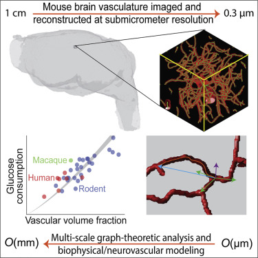 Brain microvasculature has a common topology with local differences in geometry that match metabolic loadXiang Ji, Tiago Ferreira, Beth Friedman, and 5 more authorsNeuron, Sep 2021
The microvasculature underlies the supply networks that support neuronal activity within heterogeneous brain regions. What are common versus heterogeneous aspects of the connectivity, density, and orientation of capillary networks? To address this, we imaged, reconstructed, and analyzed the microvasculature connectome in whole adult mice brains with sub-micrometer resolution. Graph analysis revealed common network topology across the brain that leads to a shared structural robustness against the rarefaction of vessels. Geometrical analysis, based on anatomically accurate reconstructions, uncovered a scaling law that links length density, i.e., the length of vessel per volume, with tissue-to-vessel distances. We then derive a formula that connects regional differences in metabolism to differences in length density and, further, predicts a common value of maximum tissue oxygen tension across the brain. Last, the orientation of capillaries is weakly anisotropic with the exception of a few strongly anisotropic regions; this variation can impact the interpretation of fMRI data.
2017
- High-Speed 4D Computational Microscopy of Bacterial Surface MotilityJaime De Anda, Ernest Y. Lee, Calvin K. Lee, and 11 more authorsACS Nano, Sep 2017
Bacteria exhibit surface motility modes that play pivotal roles in early-stage biofilm community development, such as type IV pili-driven "twitching" motility and flagellum-driven "spinning" and "swarming" motility. Appendage-driven motility is controlled by molecular motors, and analysis of surface motility behavior is complicated by its inherently 3D nature, the speed of which is too fast for confocal microscopy to capture. Here, we combine electromagnetic field computation and statistical image analysis to generate 3D movies close to a surface at 5 ms time resolution using conventional inverted microscopes. We treat each bacterial cell as a spherocylindrical lens and use finite element modeling to solve Maxwell’s equations and compute the diffracted light intensities associated with different angular orientations of the bacterium relative to the surface. By performing cross-correlation calculations between measured 2D microscopy images and a library of computed light intensities, we demonstrate that near-surface 3D movies of Pseudomonas aeruginosa translational and rotational motion are possible at high temporal resolution. Comparison between computational reconstructions and detailed hydrodynamic calculations reveals that P. aeruginosa act like low Reynolds number spinning tops with unstable orbits, driven by a flagellum motor with a torque output of \~ 2 pN μm. Interestingly, our analysis reveals that P. aeruginosa can undergo complex flagellum-driven dynamical behavior, including precession, nutation, and an unexpected taxonomy of surface motility mechanisms, including upright-spinning bacteria that diffuse laterally across the surface, and horizontal bacteria that follow helicoidal trajectories and exhibit superdiffusive movements parallel to the surface.